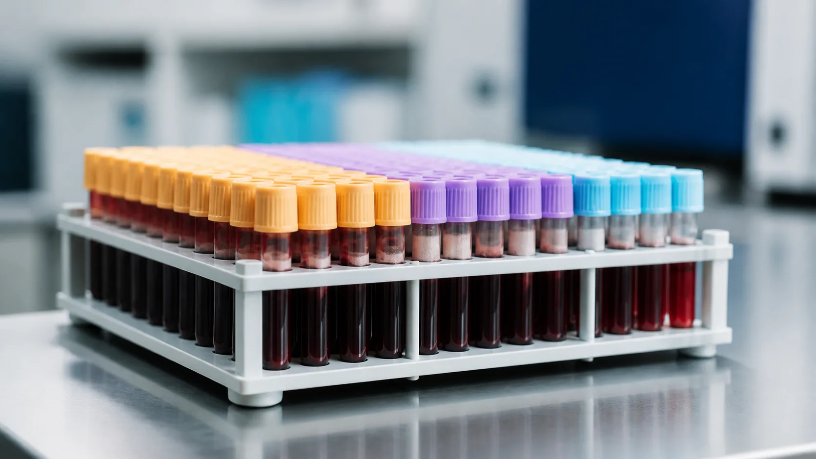
How to run a comprehensive thyroid assessment
A comprehensive thyroid assessment reads the gland on three levels at once — circulating hormones, autoantibodies and calculated structure parameters — from one venous blood sample, which means a lab requisition and a draw, not a home device. The hormones say which state you are in, the antibodies suggest why, and the calculated indices estimate reserve, conversion and central sensitivity.
What you need
One morning draw, every value run by the same laboratory from the same sample. Ask for:
- TSH — the pituitary’s signal, and the first-line test in most guidelines.
- Free T4 (free thyroxine) — the unbound fraction of the main secreted hormone.
- Free T3 (free triiodothyronine) — the active hormone, mostly made outside the gland.
- Anti-TPO and anti-Tg, the markers of autoimmune thyroiditis, plus TRAb (TSH receptor antibodies) if the pattern looks overactive.
- Your units and your lab’s reference intervals. pmol/L, ng/dL and pg/mL are not interchangeable, and the calculated parameters are unit-specific.
Conditions matter as much as the list. TSH is pulsatile and follows a circadian rhythm, so book repeats into the same morning slot, and do not test during an acute illness or just after surgery. Pause high-dose biotin. Amiodarone, lithium, glucocorticoids and estrogen shift the picture; a levothyroxine dose change needs about six weeks to settle.
How the measurement works
Take the layers in order. First the feedback loop: the pituitary releases TSH, the thyroid secretes T4 and a little T3, and circulating hormone damps the pituitary back down. That damping is roughly logarithmic, so a small drift in free T4 swings TSH a long way, which is why the two are read together. Second, the antibodies name a mechanism: anti-TPO and anti-Tg mark an autoimmune attack and usually explain an underactive pattern, while TRAb stimulate the TSH receptor instead, marking Graves’ disease.
Third, structure parameters reuse those numbers to estimate what the panel does not show directly — SPINA-GT the gland’s maximum secretory capacity, SPINA-GD the summed activity of the deiodinases that convert T4 into active T3, Jostel’s TSH index (TSHI) how strongly the pituitary responds to the free T4 in front of it, and the free T3 to free T4 ratio the same question in cruder form. These are model outputs with unit-specific constants, not arithmetic worth doing by hand; entering one set of values and getting all of them back is what a Comprehensive Thyroid Assessment does.
How to read the result
| Layer | What it answers | How to read it |
|---|---|---|
| TSH, free T4, free T3 | Which state you are in now | The combination names it: high TSH with low free T4 is overt hypothyroidism, high TSH with normal free T4 the subclinical form, the mirror images an overactive gland. |
| Anti-TPO, anti-Tg, TRAb | Whether the cause is autoimmune, and which kind | Positive or negative against an assay-specific threshold; the titer identifies the process, not its severity. |
| SPINA-GT, SPINA-GD, TSHI, free T3/free T4 | Secretory reserve, conversion, central sensitivity | As a position inside the range your calculator prints, and a direction of travel across repeat draws. |
Reference intervals are conventions, not constants. Most laboratories place the TSH upper limit near 4 mIU/L, but that figure is assay-specific, drifts upward with age and is replaced by pregnancy-specific ranges; antibody cut-offs and the ranges for the calculated parameters belong likewise to the assay they came from. An abnormal result is therefore repeated weeks later, under the same conditions, before it is acted on. Where the one question is how much hormone the gland could still make, Maximum thyroid secretory capacity takes that estimate on its own.
What it does not tell you
It does not see the gland. Nodules, goiter and thyroid cancer commonly sit behind a normal panel, so clean results do not clear a lump in the neck; that belongs to ultrasound. Antibodies carry no timetable: positive anti-TPO raises the risk of overt hypothyroidism, but many people carry them for years with normal hormones, and a negative result excludes nothing.
The calculated layer is the softest. These are interpretive research tools resting on model assumptions and a steady state; they inherit every error in the underlying immunoassays, and most have no consensus clinical thresholds — a caveat that applies wherever Thyroid hormone sensitivity is reported. During serious illness free T3 falls and TSH misbehaves as a normal adaptation, so all three layers misread at once. Biotin and interfering antibodies distort the assays themselves.
Nor does it explain symptoms or decide treatment. Fatigue, weight change and low mood are non-specific: a normal result does not mean nothing is wrong, and an abnormal one does not prove the thyroid is what you feel. No outcome trial shows that treating a calculated index helps. Where the pattern points toward hypothyroidism or its opposite, the decision belongs with a clinician reading the whole picture.
Related reading
Disclaimer. This article is for information only and does not replace medical advice. Talk to a qualified clinician before changing anything about your health.