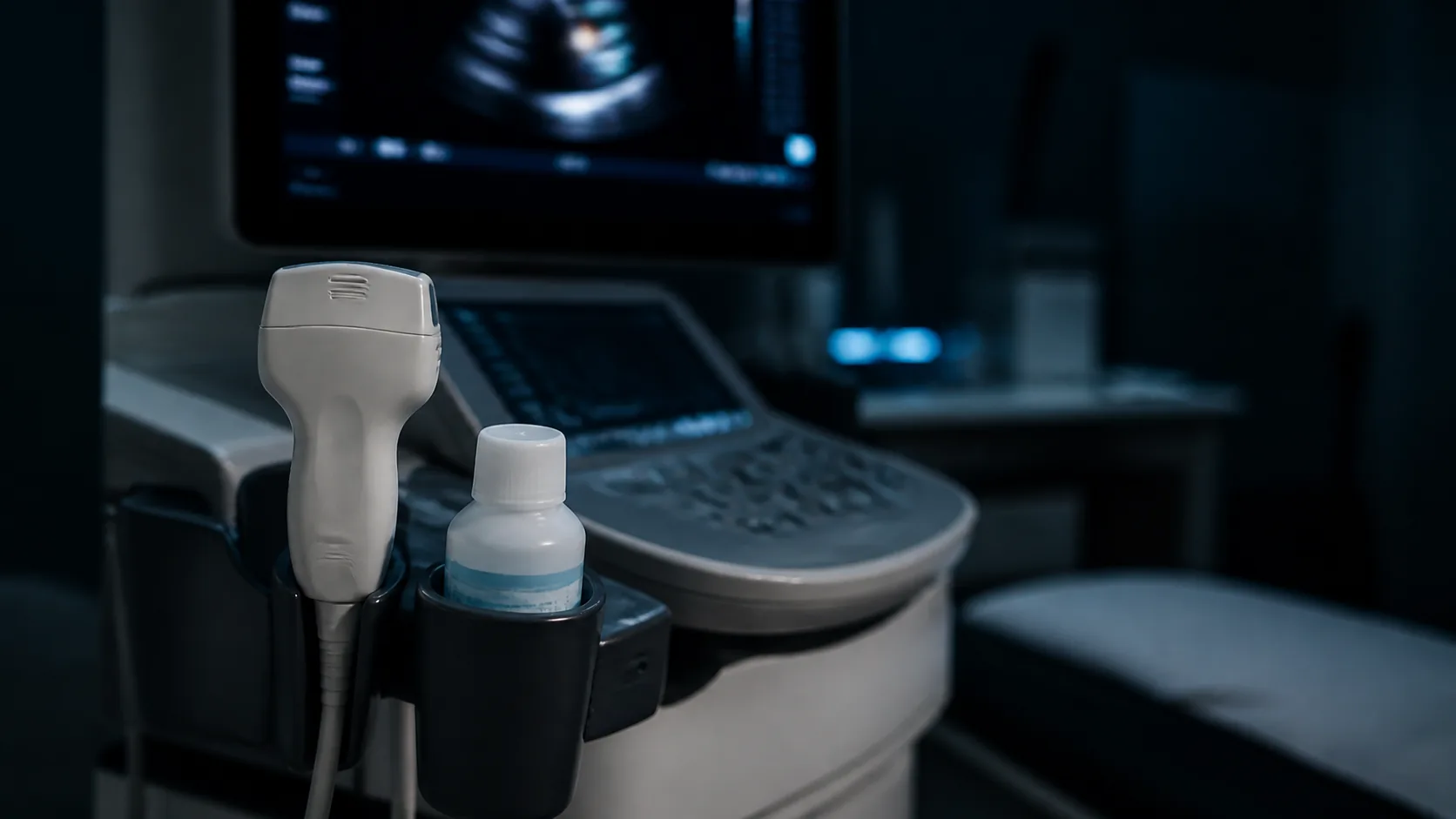
How an echocardiogram estimates your heart age
Heart age from an echocardiogram is your calendar age re-estimated from how your heart looks and moves on ultrasound — and it is not a home measurement: you first need a completed echo done in a clinic. The scan is routine: you lie on your left side, a probe is moved across the chest, and twenty to forty-five minutes later a report comes back full of millimeters, percentages and Doppler velocities. The age estimate is produced afterward, from those numbers alone.
What you need
Almost everything here comes from the scan, and in most health systems a transthoracic Echocardiography (echo) needs a physician’s referral.
- A completed transthoracic echocardiogram — no radiation, no fasting, no preparation.
- The full written report with its measurement table, not the summary line alone. “No abnormality detected” contains none of the values a model needs.
- Your age and sex, the model’s reference point.
- From the report: left ventricular wall thickness, left ventricular internal dimension or volume, left atrial size, ejection fraction, and the Doppler filling measurements (E and A waves, the E/A ratio, tissue Doppler e’ and E/e’).
- The units as printed: wall thickness appears in millimeters in some labs and centimeters in others, and a misplaced decimal ruins the result.
How the measurement works
A heart ages in a fairly predictable pattern that shows up on ultrasound. The walls of the left ventricle thicken modestly while the cavity stays the same size or shrinks a little. Early relaxation slows, so filling depends more on the atrial kick — that is what a falling e’ velocity and a falling E/A ratio describe. The left atrium tends to enlarge as filling pressure rises. Ejection fraction, the headline number most people look for, barely moves with healthy aging, which is why a normal reading there tells you little about the age of the heart.
A model is trained on many scans in which the person’s real age was known, learns which combination of measurements matches which decade, and is then given your numbers. The A cardiac ultrasound reveals your biological age calculator does that arithmetic from a small set of standard echo parameters; the value is the model’s, not something you can judge by eye.
How to read the result
The output is a number of years, read against your calendar age. There are no clinical bands and no cut-off separating healthy from unhealthy — only a direction of travel.
| Result | What it means |
|---|---|
| Below your calendar age | Measurements resembling a younger reference group |
| Roughly equal | Typical structure and filling for your age and sex |
| Above your calendar age | Thicker walls, slower filling or a larger atrium than usual for your age |
Size matters more than sign. Models of this kind report an average error of roughly five years, so a gap of one or two years is noise and only a larger, repeated gap is worth raising with a cardiologist. Without an echo report, an electrocardiogram-based estimate such as AI reveals your heart’s age is a cheaper, more available route to the same idea, though it reads electrical signals rather than structure and the two are not interchangeable.
What it does not tell you
It is not a diagnosis and does not replace the cardiologist’s reading of the same report: valve disease, wall-motion abnormalities and congenital findings live in the report text, not in the age number.
It inherits every weakness of the scan and of its training set. Echo measurements are operator-dependent: image quality, probe angle and caliper placement all shift them, and a repeat scan on another machine will not reproduce them exactly. Atrial fibrillation makes the A wave disappear and the filling measurements meaningless. Endurance athletes have larger, thicker hearts for reasons unrelated to aging, and valve replacement, a pacemaker or pregnancy move the same measurements the model reads as age.
And it is narrow. It describes the muscle and its filling, not your arteries, lipids or blood pressure, so a young result says nothing about plaque. An older result never names its cause either — hypertension, sleep apnea, alcohol, diabetes and excess weight all push these measurements the same way. It is a snapshot of the heart today, not a prediction of your future.
Related reading
- Heart Health: A Prevention Guide for Longevity
- What Biological Age Is and How to Measure It
- Vascular age
- Cardiovascular disease (CVD)
Disclaimer. This article is for information only and does not replace medical advice. Talk to a qualified clinician before changing anything about your health.